The
increasing prevalence of tattoos provoked safety concerns with respect
to particle distribution and effects inside the human body. We used skin
and lymphatic tissues from human corpses to address local biokinetics
by means of synchrotron X-ray fluorescence (XRF) techniques at both the
micro (μ) and nano (ν) scale. Additional advanced mass
spectrometry-based methodology enabled to demonstrate simultaneous
transport of organic pigments, heavy metals and titanium dioxide from
skin to regional lymph nodes. Among these compounds, organic pigments
displayed the broadest size range with smallest species preferentially
reaching the lymph nodes. Using synchrotron μ-FTIR analysis we were also
able to detect ultrastructural changes of the tissue adjacent to tattoo
particles through altered amide I α-helix to β-sheet protein ratios and
elevated lipid contents. Altogether we report strong evidence for both
migration and long-term deposition of toxic elements and tattoo pigments
as well as for conformational alterations of biomolecules that likely
contribute to cutaneous inflammation and other adversities upon
tattooing.
Introduction
In recent years, the seemingly unstoppable trend for tattoos has brought safety concerns into the spotlight1.
Currently, basic toxicological aspects, from biokinetics to possible
alterations of the pigments, are largely uncertain. The animal
experiments which would be necessary to address these toxicological
issues were rated unethical because tattoos are applied as a matter of
choice and lack medical necessity, similar to cosmetics2.
Consequently, the hazards that potentially derive from tattoos were as
yet only investigated by chemical analysis of the inks and their
degradation products in vitro3,4,5,6. Even though toxicological data might be available for some ink ingredients individually, information on in vivo interactions of the ink’s components and their fate within the body is rare.
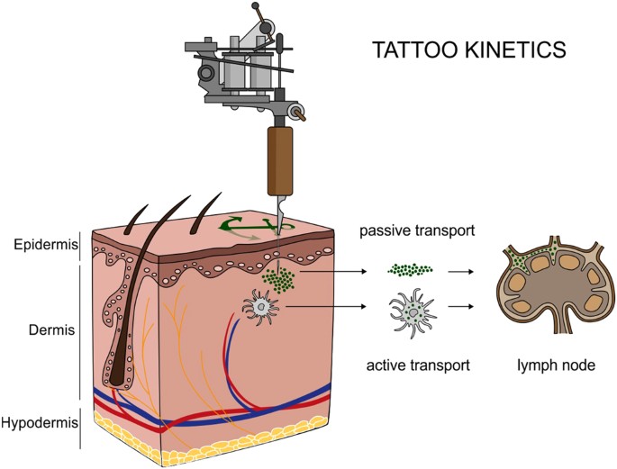
Tattoos and permanent make-up work by depositing insoluble pigments into the dermal skin layer (Fig. 1). In conjunction with tattoos, pigmented and enlarged lymph nodes have been noticed in tattooed individuals for decades7.
After the traumatic insertion of inks during the tattooing procedure,
the body will excrete as many components as possible via the damaged
epidermis. Other ways to clean the site of entrance are through active
transport to lymph nodes by phagocytizing cells, or passively along the
lymphatic vessels8,9,10,11. In addition to observations in humans, an in vivo study in mice revealed colored lymph nodes after tattooing with an azo pigment12.
Even though this leaves little doubt that the pigment originates from
corresponding tattoos, the origin and fate of pigments in human lymph
nodes have never been analytically investigated so far. Lately, the only
study analyzing human lymph nodes in tattooed individuals assessed its
contents on carcinogenic polycyclic aromatic hydrocarbons deriving from
carbon black particles13.
Figure 1
Translocation
of tattoo particles from skin to lymph nodes. Upon injection of tattoo
inks, particles can be either passively transported via blood and lymph
fluids or phagocytized by immune cells and subsequently deposited in
regional lymph nodes. After healing, particles are present in the dermis
and in the sinusoids of the draining lymph nodes. The picture was drawn
by the authors (i.e., C.S.).
Tattoo pigments consist of either inorganic
colorful metals and its oxides, or of polyaromatic compounds, all of
which are thought to be biologically inert. It can thus be expected to
find a broad range of elements in tattooed human tissue—among them the
sensitizers nickel (Ni), chromium (Cr), manganese (Mn), and cobalt
(Co)—as parts of color-giving pigments or element contamination14,15,16,17. Besides carbon black, the second most common used ingredient of tattoo inks is titanium dioxide (TiO2), a white pigment usually applied to create certain shades when mixed with colorants. The toxicity of TiO2 depends on its speciation (crystal structure) which can be either rutile or the more harmful photocatalytically active anatase18.
The latter can lead to the formation of reactive oxygen species when
exposed to sunlight. Delayed healing is thus often associated with white
tattoos, along with skin elevation and itching19.
The contribution of tattoo inks to the overall body load on toxic elements, the speciation of TiO2,
and the identities and size ranges of pigment particles migrating from
subepidermal skin layers towards lymph nodes have never been
analytically investigated in humans before. The average particle size in
tattoo inks may vary from <100 nm="" to="">1 µm20. Therefore most tattoo inks contain at least a small fraction of particles in the nano range.
Here,
we analyzed tattooed human skin and regional lymph nodes originating
from four donors (corpses). Inductively coupled plasma mass spectrometry
(ICP-MS) was used to assess the general contents of elements in both
tissues and tattoo inks after microwave digestion.
Laser-desorption/ionization time-of-flight (LDI-ToF) MS facilitated the
identification of organic pigments in enzyme-lysed samples. To precisely
locate the different elements in the cutaneous and lymphatic
microenvironments, to provide information on TiO2 speciation
and to assess primary particle sizes at the nanometric scale in particle
mixtures, however, synchrotron-based X-ray fluorescence investigations
have been performed at both the micro (μ-XRF) and nano (ν-XRF) range.
Furthermore, we assessed biomolecular changes in the respective tissues
at the micrometric scale and in the proximity of tattoo particles using
synchrotron-based Fourier transform infrared (μ-FTIR) spectroscopy.
Results
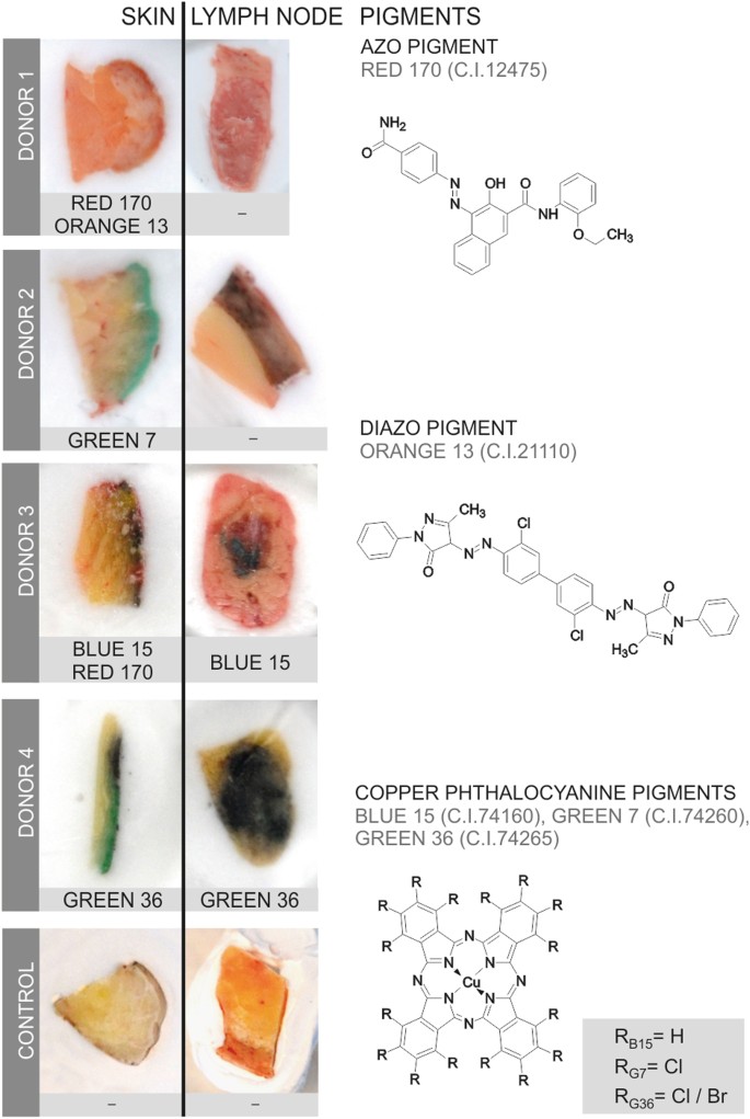
Organic pigments translocate from skin to lymph nodes
Providing
analytical evidence of tattoo particles being distributed inside the
human body was a key objective of this investigation. To this end,
tissue samples of four individuals tattooed with orange, red, green or
black and two non-tattooed control donors were analyzed for the presence
of organic pigments. Detection of the same pigment species in both skin
and regional lymph nodes revealed the drainage of tattoo particles in
two out of four tattooed donors (Fig. 2).
Figure 2
Organic
pigments translocate from skin to lymph nodes. Organic pigments in
lysed skin and lymph nodes were identified by means of LDI-ToF-MS.
Adjacent skin and lymph tissue specimens (about 5–10 mm) are displayed
in cryo-matrix after preparing thin sections for μ-FTIR and μ-XRF
analyses. Skin specimens are oriented with its surface on the right
side. Identified organic pigments are indicated below each sample.
Chemical structures of the organic pigments identified in the samples
are displayed on the right.
Identification of organic pigments using LDI-ToF-MS has mostly been described using inks21,22,23. This technique is mainly based on isotope distributions and the molecular mass (see Supplementary Fig. S1).
In the lysed tissues presented here, color-giving pigments were found
to be copper phthalocyanines with either hydrogen, chlorine or bromine
residues in three out of four skin samples. Reddish parts of the tattoos
contained the azo group-containing pigments red 170 and orange 13
(Fig. 2).
For
donors 1 and 2, the absence of organic pigments in the lymph nodes
suggests either concentrations below the limit of detection (approx.
0.1–1% w/w pigment per extract), possible metabolic decomposition or
drainage to alternative lymph nodes. The general ability for azo pigment
translocation to lymph nodes was proven in additional skin and lymph
node samples of donor 2 (Supplementary Table 1). On the other hand, carbon black particles possibly responsible for the black color in skin and lymph nodes (Fig. 2)
were not accessible with the analytical methods used in this
investigation. No xenobiotic pigment particles were detected in either
skin or lymph tissue of the control samples.
Tattoos contribute to the elemental load of lymph nodes
A
central aim of this study was to assess to what extent tattooing
increases the proportion of toxic elements in the body. We found Al, Cr,
Fe, Ni and Cu quantitatively elevated in skin and lymph node specimens
using ICP-MS analysis (Table 1 and Supplementary Table S2).
For donor 4, Cd and Hg concentrations were found increased only in the
lymph nodes, but not in the analyzed skin sections. These elements
probably result from other tattoos that were not part of this study or
other routes of exposure drained through the same lymphatic tissue.
Non-quantitative evaluation of the survey scans revealed the presence of
Ti, presumably derived from TiO2, in all tattooed skin samples but not in controls.
Table 1 Element concentrations per tissue weight (ppm) in human skin and lymph node samples analyzed by ICP-MS.
The microwave digestion used in this
investigation is not suitable to fully dissolve Fe and Ti, although no
residual particles were visible. Therefore Fe concentrations might not
represent the total amount in the samples, but they enable the
distinction between physiological concentrations in controls and samples
containing extrinsic Fe. The elevated levels of Fe found in the skin
and lymph nodes of donor 4 imply an additional use of iron-based
pigments. In donors 1, 2 and 3, Fe concentrations were only increased in
adjacent lymph nodes and not in the corresponding skin samples (Table 1). Fe concentrations can also be affected by residual blood within the tissue samples.
In
donor 4, the use of pigment copper phthalocyanine green 36, as
identified with LDI-ToF-MS, is reflected by high amounts of Cu in skin
and lymph nodes as well as the non-quantitative detection of Br (Table 1).
By contrast, although pigment copper phthalocyanine green 7 was well
detectable with our LDI-ToF-MS approach in the skin of donor 2, it was
not in the corresponding regional lymph node. Increased Cu levels
measured by ICP-MS in this adjacent sample, however, suggest the
presence of this copper phthalocyanine pigment. In light of the other
two copper phthalocyanines applied in donor 2 (green 7) and 3 (blue 15)
elevated Cu levels in skin came without surprise (Table 1).
In donor 2, Cu levels in lymph nodes are strongly increased despite the
fact that green 7 could not be detected with LDI-ToF-MS. However,
adjacent samples of tissue were used for each analysis. Given the nature
of the samples, pigment deposition within skin and lymph nodes is not
homogeneous and therefore explaining the different findings.
Interestingly, the non-tattooed control donor 1 also had slightly
elevated levels of about 13 ppm Cu in the lymph nodes which is still in
the range of the average 5.89 ± 8.03 ppm of Cu detectable in lymph nodes
of female cadavers (Table 1)24.
Additionally,
Ni and Cr were found in the human specimens. Since Ni levels were
increased in the skin and lymph nodes of donor 2 and 3, the likely
source is the tattoo. In different studies, both elements were linked to
adverse reactions occurring in tattooed patients25,26,27,28.
Ni and Cr are known to be allergenic as well as carcinogenic. Ni
concentrations of 0.28–10.05 ppm total tissue weight found here are
within the range of 0.8–3.7 ppm dry weight Ni in hilar lymph nodes in
previous studies29.
Cd was drastically elevated only in the lymph node of donor 4. For all
other samples, Cd tissue burdens lie within normal values24.
Finally, Al was also present in skin and lymph node tissues of the three tattooed donors 2, 3 and 4 (Table 1).
Since auxiliary lymph nodes have been investigated in the case of donor
2 and 3, co-exposure from antiperspirants containing various aluminum
salts cannot be excluded, neither in tattooed nor control samples.
However, Al concentrations in the controls were lower. The light metal
Al has recently attracted attention because of its accumulation in
breast cancer tissue30.
While its role in the emerging of neoplasia is currently highly
disputed, its contribution to the occurrence of hypersensitivity
granulomas associated with tattoos has been proven since decades31.
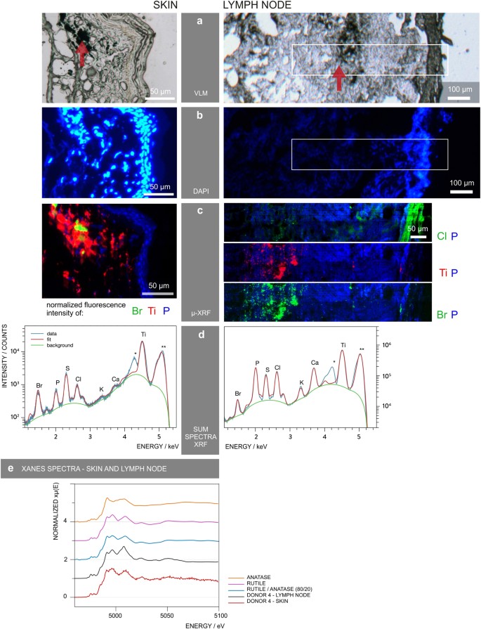
μ-XRF mapping links metallic elements to tattoo particles
In
order to link elements found with ICP-MS in tattoo pigment particles
and to locate them inside the tissues, μ-XRF imaging was carried out
with sub-micrometric probes over skin and lymph node sections (Fig. 3a–d).
The location of particles can be altered by sample preparation. Since
transversal sections were made by moving the knife parallel to the skin
surface, the depth profile of the pigments should remain unaffected.
Thin sections of skin and lymph nodes from donors 1, 3 and 4 were
analyzed at the ESRF beamline ID21, with an exciting energy of 5.05 keV
(Fig. 3a–d and Supplementary Fig. S2). Since the thin sections were deposited on BaF2
windows for further μ-FTIR analyses, the energy was chosen to avoid
excitation of Ba L-lines (<5 .24="" 4="" a="" are="" data-track-action="figure anchor" data-track-label="link" data-track="click" displayed="" donor="" fig.="" href="https://www.nature.com/articles/s41598-017-11721-z#Fig3" in="" kev="" nbsp="" of="" results="">3
as an example.
Figure 3
μ-XRF
mapping identifies and locates tattoo particle elements in skin and
lymph node tissue sections. Sections of skin and lymph node tissue from
donor 4 were analyzed by means of synchrotron μ-XRF. (a) Visible light microscopy (VLM) images of the area mapped by μ-XRF. Tattoo pigments are indicated by a red arrow. (b) DAPI staining of adjacent sections showing the cell nuclei. (c) μ-XRF maps of P, Ti, Cl and/or Br. For the lymph node, areas of similar size are marked in (a) and (b). (d) Average μ-XRF spectra over the full area displayed in (c) *diffraction peak from sample support; **scatter peak of the incoming beam. (e)
Ti K-edge μ-XANES spectra of skin and lymph node compared to
transmission XANES spectra of reference material of rutile, anatase and
an 80/20 rutile/anatase mixture calculation.
The majority of particles in the skin tissue
were surrounded by phosphor-rich nuclei visualized by DAPI staining in
fluorescence light microscopy (Fig. 3b) and integration of the element P in μ-XRF analysis (Fig. 3c). It was previously shown that tattoo particles can primarily be found around vessels10 which might account for the high cell density in the dermis co-localized with the pigments.
Intensities of Ti K-lines and Br L-lines were extracted to map the distribution of TiO2 and the highly brominated pigment copper phthalocyanine green 36 (Fig. 3c).
Since the Br L-lines completely overlap with the Al K-lines, both may
contribute to the intensity of the peak. However, LDI-ToF-MS analysis
revealed the presence of pigment green 36 (Fig. 2)
and the following ν-XRF results from ID16B acquired at 17.5 keV, i.e.
above Br K-edge (13.47 keV) undermined the primarily Br-related
contribution (see Supplementary Fig. S3).
Tattoo
particles containing Ti and Br are adjacent to each other with only a
slight overlap in skin and seem to be more evenly co-localized in lymph
tissue (Fig. 3c).
Both elements were found in the dermis of donor 4 directly beneath the
cell nuclei-rich epidermis and up to a few hundred micrometers deep in
the skin. In the lymph nodes, some particles were deposited in the
stroma directly beneath the capsule. The bulk of Ti and Br containing
particles, however, became visible as pigment agglomerates at a distance
of about 250 µm to the lymph node capsule. Conversely, Cl
concentrations are highest in the lymph node capsule and lower
concentrations can be found in the particle region as part of the
pigment phthalocyanine green 36.
All analyzed samples from the
tattooed donors contained Ti. It is unlikely that other sources, e.g.
sun screens, would explain the high amounts found in this investigation.
Elevated amounts of Ti are only expected in lung and hilar lymph nodes
from respiratory exposure32. Other highly abundant elements are K and Ca as they are physiologically present in cells (Fig. 3d).
We also investigated if the Ti present is the expected white pigment TiO2
and whether the stable rutile and/or the more photoreactive anatase
crystal phases were used in tattoo inks. Micro X-ray absorption near
edge structure (μ-XANES) spectra at the Ti K-edge were collected for the
skin and lymph nodes of donors 1, 3 and 4. The spectra of donor 4
showed more qualitative correlation with the reference spectrum of
rutile than with that of anatase (Fig. 3e).
A clear switch of peak maxima between 4.99–5 keV occurs as a difference
of both types of crystal structures. A calculated spectrum of 20%
anatase and 80% rutile mixture is not clearly distinguishable from pure
rutile, but shows a pre-edge at around 4.97 keV, similar to the μ-XANES
spectra of the tattooed samples. Therefore, mostly rutile TiO2 is present in all tattooed donors, with minor amounts of anatase (Fig. 3e and Supplementary Fig. S2).
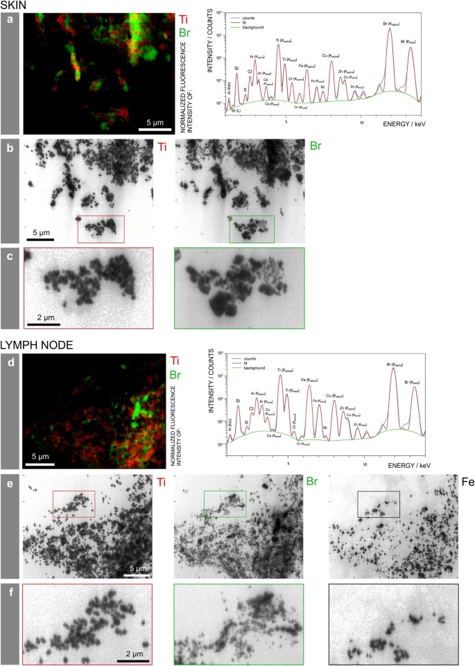
Particle size varies between pigment species
The obtained μ-XRF maps of skin and lymph node sections show large tattoo particle agglomerates up to several micrometers (Fig. 3c).
However, it is known that small-sized particles are preferentially
transported to lymph nodes. The 0.3 × 0.7 µm² beam size for μ-XRF
mapping at ID21 was therefore a limiting factor for the determination of
particle sizes. To assess the primary particle sizes, we additionally
performed ν-XRF investigations by applying a beam of 50 × 50 nm² at
17.5 keV in order to excite the Br K-lines. Experiments were carried out
in adjacent sections of skin and lymph node from donor 4, prepared on
ultralene foil (Fig. 4).
We detected three different pigment particles, each showing a different
elemental composition and distribution within the same area (Fig. 4b,e). The average particle size of TiO2
in both skin and lymph nodes was 180 nm with a standard deviation of
23 nm and a standard error of 7 nm. Therefore this rather large particle
size does not prevent distribution via the lymph fluid.
Figure 4
Particle
mapping and size distribution of different tattoo pigment elements.
Skin and lymph node of donor 4 were analyzed by means of synchrotron
ν-XRF. (a,d) Ti and the Br containing pigment
phthalocyanine green 36 are located next to each other. Average XRF
spectra over the full area displayed in the regions of interest reveal
the presence of Br, Si, S, Cl, Ca, Ti, Cr, Fe, Ni, Cu, and Zn. (b,e) Log scale mappings of Ti, Br and Fe in the same areas as displayed in (a) and (d) reveal primary particle sizes of different pigment species. (c,f) Magnifications of the indicated areas in (b) and (e), respectively.
In contrast, the pigment phthalocyanine green
36 analyzed by ν-XRF mapping of Br was much more polydisperse, with
particles presumably smaller than the resolution of 50 nm and up to the
µm range in skin. In lymph node tissue, particles containing Br were
smaller, with fewer particles of a larger size (Fig. 4c,f). Hence it can be assumed that the transport of smaller particles is preferential.
With
the chosen energy, Br can be unequivocally identified from its K-lines
emission. The skin and lymph node of donor 4 also contained Cu, related
to the identified copper phthalocyanine pigments, and its maps show
perfect co-localization with Br (see Supplementary Fig. S3).
Additionally, Fe particles were present in the lymph node but not skin
tissue and therefore possibly originate from another tattoo or route of
exposure (Fig. 4c,f and Supplementary Fig. S3).
Tattoo particles induce biomolecular changes
The
synchrotron-based μ-FTIR end-station at ID21 was used to monitor
changes in protein conformation as well as in the overall protein and
lipid contents in the proximity of tattoo particles. Synchrotron μ-FTIR
analyses allow the assumption that tattoo pigments became located in a
lipid-rich β-sheet protein environment.
The very same sections
investigated by means of μ-XRF at ID21 were analyzed by means of μ-FTIR,
prior to X-ray analyses, to facilitate exact site matching (cf.
Figures 3 and 5).
Thus, μ-FTIR results were not altered by μ-XRF radiation of the tissue
sections. The high synchrotron photon flux allowed for high spatial
resolution. Accordingly, the beam and pixel sizes were reduced to
10 × 10 µm² and 8 × 8 µm², respectively. This resolution is sufficient
to distinguish regular dermis from pigment containing areas in the
dermis, but remains insufficient to unambiguously separate the stratum corneum
from the epidermis, which were analyzed here as a single domain (see
below). Specific spectral changes related to the modification of
biomolecule composition and conformation are displayed using donor 4 as
an example, on the basis of two μ-FTIR maps obtained in a single section
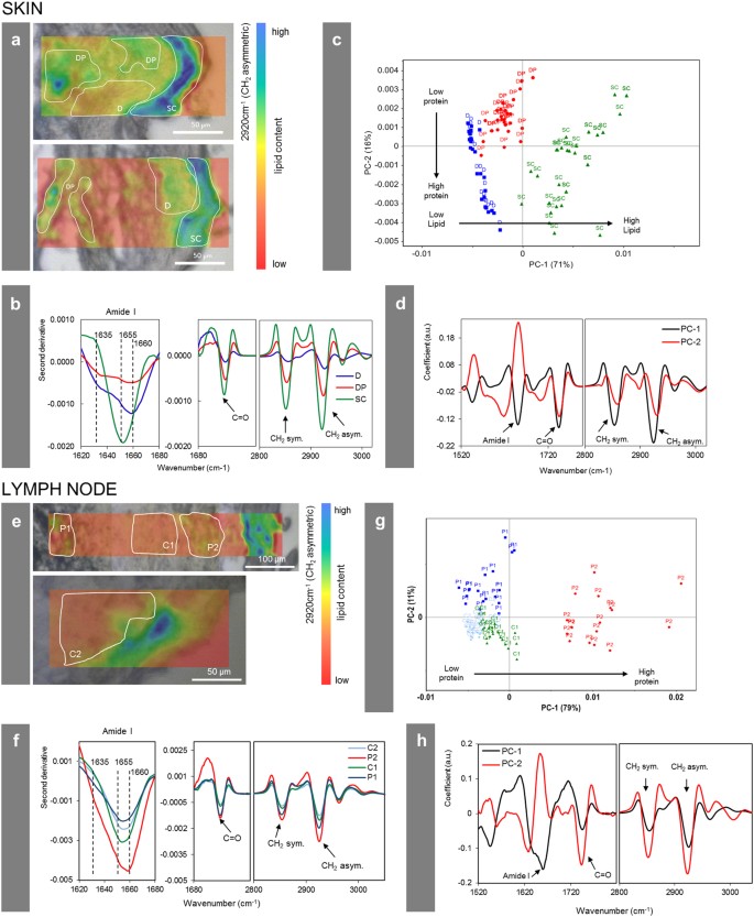
at two different locations, for the skin and regional lymph node (Fig. 5). The absorption band which peaks at 2920 cm−1 corresponds to the –CH2
stretching mode, which is much more intense in lipids than in proteins.
It can be used to qualitatively map the distribution of lipids over
thin sections (Fig. 5a). It shows a higher intensity in the stratum corneum, as expected33.
These maps also qualitatively show a higher intensity in the areas of
dermis containing tattoo pigments compared to pigment-free control
regions. Based on the microscopic images and the μ-XRF maps described
earlier, three regions were selected on each map. For the skin section,
we divided the obtained map into stratum corneum and epidermis (SC), dermis without pigment (D) and dermis around pigment particles (DP) (Fig. 5a).
Spectra in the second derivative of these areas were statistically
analyzed by means of Principal Component Analysis (PCA). Distribution of
points along the PC-1 axis confirms that D and DP have fewer
lipid-related long alkyl chains (–CH2 stretching mode, asym. at 2920 cm−1 and –CH2 sym. at 2854 cm−1) and ester (–C = O stretching mode, peak at 1745 cm−1) vibrations than SC, and that DP regions contain higher levels of lipids than D (Fig. 5b–d). PC-2 separates DP from D and SC since the latter two have higher protein concentrations.
Figure 5
Changes
of the biological composition and structure in the cellular proximity
of tattoo pigment particles. Section of donor 4 analyzed by means of
synchrotron μ-FTIR at ID21, ESRF. (a,e) Maps in second derivative obtained at 2920 cm−1 (–CH2
asymmetric vibration) of two different areas in either the skin or
lymph node of donor 4 in overlay with a visible light microscopy image.
Single points for PCA analysis in (c) and (g) were picked from the indicated areas. (b,f) Mean spectra from each region marked in (a,e) in second derivative. (c,g) PCA score plot of PC-1 vs. PC-2. (d,h) Loading plots of PC-1 and PC-2. Abbreviations: SC = stratum corneum
and epidermis; D = dermis; DP = dermis with particles; P1,
P2 = particle-containing regions; C1, C2 = control regions without
particles.
In addition to determining the component
distribution, μ-FTIR can be used to identify and map the protein
secondary structures across skin sections33. In the epidermis, keratinocytes differentiate to finally form the dead, protein- and lipid-rich stratum corneum. In the designated SC area of our investigation—comprising also the epidermal layer—the amide I peak maximum at ~1655 cm−1 (Fig. 5b) derives from α-helices present in keratin33. In the dermis, the peak maximum located at 1660 cm−1 corresponds to triple helices present in collagen, while the β-sheet shoulder at 1635 cm−1 can be assigned to crosslinked collagen fibers33.
In the proximity of the pigment particles (DP), the protein content is
lower compared to other parts of the collagen-rich dermis. However, the
β-sheet shoulder at 1635 cm−1 becomes more pronounced close to the particles (Fig. 5b). The –CH2 and –C = O
vibrations related to lipids are also higher in the proximity of
particles compared to other parts of the dermis. Both findings suggest
the presence of denatured β-sheet-rich protein and lipid membranes
surrounding the pigment particles. Other investigations have shown that
when in contact with foreign surfaces, protein structures can be altered
towards the formation of β-sheets34.
In the skin of donor 2, a similarly enhanced lipid content and the
presence of β-sheet structures in the dermis around particles were also
noticed (see Supplementary Fig. S4).
A statistical comparison of particle-containing and particle-free areas
in the lymph node tissue of donor 4 showed a similar increase of lipid
contents in the former (Fig. 5e–h). However, no consistent differences in the kind of protein folding could be observed in lymph nodes.
Discussion
In
this investigation, we found a broad range of tattoo pigment particles
with up to several micrometers in size in human skin but only smaller
(nano)particles transported to the lymph nodes. The exact size limit
preventing this translocation is unknown yet. The deposit of particles
leads to chronic enlargement of the respective lymph node and lifelong
exposure. With the detection of the same organic pigments and inorganic
TiO2 in skin and lymph nodes, we provide strong analytical
evidence for the migration of pigments from the skin towards regional
lymph nodes in humans. So far, this only has been assumed to occur based
on limited data from mice and visual observations in humans13, 35.
We also were able to prove the presence of several toxic elements, such
as Cr and Ni, derived from tattooing. However, elemental deposits in
lymph nodes which were not found in the corresponding skin revealed that
tattooing might not have been the only route of exposure in these
particular individuals whose tissues were removed after their demise.
It is known that pigments reside in lysosomes or stay attached to membranes of dermal fibroblasts8, 36,
an observation that supports our μ-FTIR findings of concentrated lipid
levels in the proximity of pigment particles. Long alkyl chains and
ester groups which we assigned to lipids may also derive from components
of tattoo inks, e.g. thickening polymers, surfactants and pigment
coatings. However, the frequently used polyethylene glycol37 and polyvinyl pyrrolidone polymers below 20 kDa38
are known to be metabolized and secreted. In addition, the strong
lysosomal and reactive oxygen species-driven reaction of macrophages
against foreign material was shown to alter even the highly stable
polyurethane39. We therefore assume these additives being biodegradable in vivo
and thus not anymore present in healed tattooed skin. Since the
initially used coatings and surfactants of the particles were unknown,
interferences in μ-FTIR cannot be fully excluded though.
In cases
where foreign hydrophobic material is introduced into the body,
fibrinogen and other proteins are likely to become adsorbed and
denatured at its surface, thus leading to the generation and
presentation of pro-inflammatory matter and subsequent recruitment of
immune cells as the initial step in the triggered foreign body reaction40, 41.
This assumption becomes supported by our μ-FTIR data on β-sheet
associated conformational changes of proteins in the proximity of
hydrophobic, insoluble tattoo pigments. Foreign body reactions are known
from subcutaneous injections of TiO242.
Despite the hydrophobic nature of pigment surfaces and the here
confirmed β-sheet protein conformation in the proximity of tattoo
pigments in skin, most tattooed individuals including the donors
analyzed here do not suffer from chronic inflammation though. Yet,
granulomatous foreign material reactions are among the main
non-infectious side effects occurring upon tattooing43.
Factors preventing the progression towards adverse foreign body
reactions in most tattooed individuals despite a β-sheet conformation
need to be further investigated.
In future experiments we will
also look into the pigment and heavy metal burden of other, more distant
internal organs and tissues in order to track any possible
biodistribution of tattoo ink ingredients throughout the body. The
outcome of these investigations not only will be helpful in the
assessment of the health risks associated with tattooing but also in the
judgment of other exposures such as, e.g., the entrance of TiO2 nanoparticles present in cosmetics at the site of damaged skin.
Methods
Human sample preparation
Samples
of tattooed skin and regional lymph nodes as well as skin and lymph
node samples of two additional donors without any tattoos were taken postmortem
at the Institute of Forensic Medicine at the Ludwig-Maximilians
University of Munich (court-ordered autopsies without any additional
cosmetic impairment to the skin). The experiments were performed
according to the Helsinki Declaration of 1975. All samples were obtained
anonymously without information on the patients disease status, causes
of death or demographies. Ethical approval of human biopsy samples was
granted by the Ethics committee of the Medical Faculty of the
Ludwig-Maximilians University of Munich. We selected specimens with
tattoos other than black and which are more likely to contain TiO2
and organic pigments. The sample size was limited by the availability
of specimens and the beamtime at ESRF. Tissue samples were stored in
plastic bags at −20 °C directly after excision and further processed for
analysis within a year. Subsamples were cut using a standard scalpel
and frozen in TissueTek O.C.T. matrix (Sakura Finetek, Staufen, Germany)
for cryo-microtome sectioning. Sections of 5 or 6 µm were obtained and
mounted on BaF2 substrates (Crystal GmbH, Berlin, Germany)
for μ-FTIR and μ-XRF measurements at ID21. Sections for fluorescence
light microscopy had a thickness of 6–10 µm and were deposited on
standard glass slides, while ν-XRF analyses at ID16B were performed on
12–14 µm sections on 4 µm Ultralene window films (Spex Sample Prep,
Metuchen, NJ, USA) mounted on Si3N4 windows.
Sections were inactivated using 4% formaldehyde buffer for 10 min and
subsequently washed with deionized water (2 times, 2–5 min). For μ-FTIR
and μ-XRF analyses, samples were freeze-dried and stored in a dehydrated
environment. Sections on microscopic glass slides were mounted in
DAPI-Fluoromount G (Southern Biotech, Birmingham, AL, USA) for cell
nucleus staining.
ICP-MS analysis
Elemental
compositions of in total 20 skin and 25 lymph node samples of tattooed
donors as well as 2 skin and 2 lymph node samples of non-tattooed donors
were analyzed using a nitric acid microwave digestion (Ultraclave, MLS,
Leutkirch, Germany). Samples were directly adjacent to those used in
other parts in this investigation. Five milliliter of 69% nitric acid
was added to 50–200 mg tissue samples in Teflon vessels and heated in
the microwave with the following steps: 20–80 °C (3.5 min, 100 bar,
700 W); 80–130 °C (10 min, 120 bar, 1000 W); 130–200 °C (6.5 min,
150 bar, 1000 W), 200 °C (30 min, 150 bar, 1000 W). Elemental
concentrations given in ppm are calculated in relation to the weight of
digested tissue. Nitric acid was purified using a duoPUR quartz
sub-boiling distillation system (MLS, Leutkirch, Germany). Ultrapure
water was obtained using a Milli-Q Advantage A10 water purification
system equipped with a Millipore Q-POD Element Unit (both from Merck,
Darmstadt, Germany). Standards for ICP were purchased either from Sigma
Aldrich (Munich, Germany; i.e. Sc, Al, Cu, Ni, Hg) or Merck (Darmstadt,
Germany) in the case of In. For Cr, Fe and Cd 1000 mg/l standard
solutions in diluted nitric acid were obtained from VWR (Darmstadt,
Germany).
A 20-fold dilution of each sample was prepared including
10 ppb of the elements In and Sc as internal standards. XSeries II
ICP-MS (Thermo Fischer Scientific, Bremen, Germany) together with an ESI
SC2 autosampler (Elemental Service & Instruments, Mainz, Germany)
were used for sample analysis. Sample analysis was carried out in
triplicate with 100 sweeps each. Resolution was set to 0.02 amu and the
dwell time for all elements was 10 ms. Measurements were carried out
with collision cell in either −3.0 V mode (In, Sc, Cr, Fe, Ni, Cu, Cd)
or 0.0 V mode (Sc, Al). H2/He (7% v/v) was used as the
collision gas with 5 ml/min flow rate. Data were processed with
PlasmaLab 2.5.11.321 (Thermo Scientific, Bremen, Germany).
LDI-ToF-MS identification of organic pigments
In
total 8 skin and 8 lymph node samples of tattooed donors as well as 2
skin and 2 lymph node samples of non-tattooed donors were analyzed.
Samples between 50–200 µg were lysed using 1 mg/ml collagenase from Clostridium histolyticum
Type IA (Sigma Aldrich, Munich, Germany) with an incubation time of at
least 24 hours at 37 °C. Lysates were heat-inactivated at 90–95 °C for
at least 12 hours. Precipitated pigment particles were washed twice with
PBS. Centrifugation was carried out with 500× g for 10 min.
Precipitates were applied as thin films to a ground steel target plate
with a plastic pipette tip and measured using an UltrafleXtreme
MALDI-ToF/ToF (Bruker Daltonik, Bremen, Germany). Spectra were obtained
by averaging 3000 individual spectra, with a laser rate of 1000 Hz in
positive reflector mode. The instrument was calibrated prior to each
measurement with an external ProteoMass™ MALDI Calibration Kit (Sigma
Aldrich, Munich, Germany). Data were processed using the flexControl 3.4
and flexAnalysis 3.4 software (Bruker Daltonik, Bremen, Germany).
Synchrotron FTIR microscopy
FTIR
microscopy analyses were performed at beamline ID21 at the European
Synchrotron Radiation Facility (ESRF) in Grenoble, France44.
The beamline is equipped with a Thermo Nicolet Continuum (Thermo
Scientific, Madison, WT, USA) microscope coupled to a Thermo Nicolet
Nexus FTIR spectrometer (Thermo Scientific, Madison, WT, USA) with a 32x
objective, a motorized sample stage, and a liquid nitrogen-cooled 50 µm
HgCdTe detector. Maps were acquired in transmission mode using a
10 × 10 µm² beam, step size of 8 µm. Spectra were recorded as an average
of 64 scans per spectrum, over a range of 4000 to 850 cm−1 and with a spectral resolution of 4 cm−1.
The
OMNIC software was used to transform spectra from maps of skin and
lymph node samples to second derivatives using Savitsky-Golay of second
polynomial order with 21 smoothing points45, 46.
Unscrambler X software (Version 10.3, CAMO Software, Oslo, Norway) was
used for further statistical analysis. Principal component analysis
(PCA) was performed on the mean-centered data using the spectral regions
from: 1800 to 1350 cm−1 (related to proteins) and 3200 to 2800 cm−1 (related to lipids)47, 48.
PCA was performed separately for skin and lymph node samples. Score
plots and loading plots obtained by PCA analysis as well as mean values
from the regions of interest were used for data interpretation.
Synchrotron μ-XRF and μ-XANES
μ-XRF and μ-XANES analyses were carried out at the beamline ID2149.
Here, X-rays were generated by an U42 undulator operated in
“gap-tracking” mode, i.e. the gap value was optimized for each energy. A
fixed exit double-crystal Si(111) Kohzu-monochromator was used in
combination with a Ni-coated flat double-mirror rejecting high-energy
harmonics and allowed for energy selection with about 0.4 eV resolution
of the primary radiation at Ti K-edge (5.1 keV). Downstream of the
monochromator, the beam was focused down to 0.4 × 0.8 µm2 (vertical × horizontal) using a fixed-curvature Kirkpatrick-Baez (KB) mirror system. The flux was 1.6 × 10¹⁰
photons/s (~180 mA SR current in multi-bunch mode). A 30 µm Al
attenuator was used to reduce the photon flux by one order of magnitude
to keep the XRF detector dead time within its linear range. A photodiode
collecting the XRF from a thin Si3N4 membrane
inserted in the beam path was used to continuously monitor the incoming
beam intensity. XRF and scattered radiation were collected with a
dispersive energy silicon drift detector with an active area of 80 mm²
(Bruker Daltonik, Bremen, Germany). Acquisition time per point was 100
ms. The pixel size for collecting the XRF maps was adjusted to the
regions of interest and varied from 0.5 µm to 5 µm. Scans were performed
in continuous (zap) mode and an energy of 5.05 keV was selected for
μ-XRF mapping. For collecting Ti XANES spectra, the energy of the
incoming beam was scanned from 4.95 to 5.1 keV in increments of 0.5 eV,
with acquisition times of 100 ms per energy. Depending on the
concentration of the probed region, between 1 and 10 μ-XANES spectra
were collected per point and subsequently averaged. Full-field XANES
maps were also collected to total the XANES spectra over multiple
pixels.
Synchrotron ν-XRF
The
analysis on an adjacent section of skin and lymph node tissue from
donor 4 was performed by means of ν-XRF at ID16B at the ESRF. The
experimental set-up is described elsewhere50. A pink beam with an energy of 17.5 keV with ΔE/E = 1% was focused down to 50 × 50 nm² using KB mirrors. The flux of >1 × 10¹¹
photons/s was subsequently reduced using gold and silicon attenuators
to keep the dead time on the XRF detectors within the linear range. Two
three-element silicon drift detector arrays (SGX Sensortech,
Buckinghamshire, UK) were used. The two ν-XRF maps were recorded with a
step size of 50 × 50 nm² and 100 ms dwell time. In contrast to the
set-up installed at ID21, ID16B operates in air. For estimating the
particle size of TiO2, analysis was performed on 10 particles by computing the full width at half maximum of line profiles through the particles.
Availability of materials and data
XRF and FTIR data sets can be provided by the authors upon individual request.
Ethical approval of human biopsy samples
Samples of tattooed skin and regional lymph nodes were taken postmortem
and anonymously at the Institute of Forensic Medicine at the
Ludwig-Maximilians University of Munich in the frame of court-ordered
autopsies without information on the patients disease status, causes of
death or demographies. Experiments were performed according to the
Helsinki Declaration of 1975 (see: http://www.wma.net/en/30publications/10policies/b3/17c.pdf).
Ethical approval of human biopsy retrieval was granted by the Ethics
committee of the Medical Faculty of the Ludwig-Maximilians University of
Munich, Germany (confirmation by R.P., member of the ethics committee).
References
1.
Laux, P. et al. A medical-toxicological view of tattooing. The Lancet387, 395–402 (2016).
Schreiver,
I., Hutzler, C., Laux, P., Berlien, H. P. & Luch, A. Formation of
highly toxic hydrogen cyanide upon ruby laser irradiation of the tattoo
pigment phthalocyanine blue. Sci. Rep.5, 12915 (2015).
Schreiver,
I., Hutzler, C., Andree, S., Laux, P. & Luch, A. Identification and
hazard prediction of tattoo pigments by means of pyrolysis—gas
chromatography/mass spectrometry. Arch. Toxicol.90, 1639–1650 (2016).
Vasold, R. et al. Tattoo pigments are cleaved by laser light—the chemical analysis in vitro provide evidence for hazardous compounds. Photochem. Photobiol.80, 185–190 (2004).
Anderson, L. L., Cardone, J. S., McCollough, M. L. & Grabski, W. J. Tattoo pigment mimicking metastatic malignant melanoma. Dermatol. Surg.22, 92–94 (1996).
Taylor,
C. R., Anderson, R. R., Gange, R. W., Michaud, N. A. & Flotte, T.
J. Light and electron microscopic analysis of tattoos treated by
Q-switched ruby laser. J. Invest. Dermatol.97, 131–136 (1991).
Zelickson, B. D. et al. Clinical, histologic, and ultrastructural evaluation of tattoos treated with three laser systems. Lasers Surg. Med.15, 364–372 (1994).
Ferguson,
J. E., Andrew, S. M., Jones, C. J. P. & August, P. J. The
Q-switched neodymium: YAG laser and tattoos: a microscopic analysis of
laser-tattoo interactions. Br. J. Dermatol.137, 405–410 (1997).
Gopee, N. V. et al. Response of mouse skin to tattooing: use of SKH-1 mice as a surrogate model for human tattooing. Toxicol. Appl. Pharmacol.209, 145–158 (2005).
Lehner, K. et al.
Black tattoos entail substantial uptake of genotoxic polycyclic
aromatic hydrocarbons (PAH) in human skin and regional lymph nodes. PLoS One9, e92787 (2014).
Davis, M. D. et al. Patch testing with a large series of metal allergens: findings from more than 1,000 patients in one decade at Mayo Clinic. Dermatitis22, 256–271 (2011).
Dirks, M. Making innovative tattoo ink products with improved safety: possible and impossible ingredients in practical usage. Curr. Probl. Dermatol.48, 118–127 (2015).
Brady,
B. G., Gold, H., Leger, E. A. & Leger, M. C. Self-reported adverse
tattoo reactions: a New York City Central Park study. Contact Dermatitis73, 91–99 (2015).
Soltzberg,
L. J., Hagar, A., Kridaratikorn, S., Mattson, A. & Newman, R.
MALDI-TOF mass spectrometric identification of dyes and pigments. J. Am. Soc. Mass. Spectrom.18, 2001–2006 (2007).
Boon,
J. J. & Learner, T. Analytical mass spectrometry of artists’
acrylic emulsion paints by direct temperature resolved mass spectrometry
and laser desorption ionisation mass spectrometry. J. Anal. Appl. Pyrolysis64, 327–344 (2002).
Saltzman,
B. E., Gross, S. B., Yeager, D. W., Meiners, B. G. & Gartside, P.
S. Total body burdens and tissue concentrations of lead, cadmium,
copper, zinc, and ash in 55 human cadavers. Environ. Res.52, 126–145 (1990).
Morales-Callaghan,
A. M. Jr., Aguilar-Bernier, M. Jr., Martinez-Garcia, G. &
Miranda-Romero, A. Sarcoid granuloma on black tattoo. J. Am. Acad. Dermatol.55, 71–73 (2006).
Rezuke, W. N., Knight, J. A. & Sunderman, F. W. Reference values for nickel concentrations in human tissues and bile. Am. J. Ind. Med.11, 419–426 (1987).
Teraoka, H. Distribution of 24 elements in the internal organs of normal males and the metallic workers in Japan. Arch. Environ. Health36, 155–165 (1981).
Gross, N. et al.
High spatial resolution study of human skin using synchrotron infrared
microscopy: application to the penetration of external agents. IFSCC Magazine7, 1–8 (2004).
Lenk,
T. J., Horbett, T. A., Ratner, B. D. & Chittur, K. K. Infrared
spectroscopic studies of time-dependent changes in fibrinogen adsorbed
to polyurethanes. Langmuir7, 1755–1764 (1991).
Engel, E. et al. Tattooing of skin results in transportation and light-induced decomposition of tattoo pigments―a first quantification in vivo using a mouse model. Experim. Dermatol.19, 54–60 (2010).
Zhao, Q. et al. Foreign-body giant cells and polyurethane biostability: in vitro correlation of cell adhesion and surface cracking. J. Biomed. Mater. Res.25, 177–183 (1991).
Lu, D. R. & Park, K. Effect of surface hydrophobicity on the conformational changes of adsorbed fibrinogen. J. Colloid Interfac. Sci.144, 271–281 (1991).
Umbreit, T. H. et al.
Tissue distribution and histopathological effects of titanium dioxide
nanoparticles after intravenous or subcutaneous injection in mice. J. Appl. Toxicol.32, 350–357 (2012).
Wenzel,
S. M., Rittmann, I., Landthaler, M. & Bäumler, W. Adverse reactions
after tattooing: review of the literature and comparison to results of a
survey. Dermatology226, 138–147 (2013).
Martin, F. L. et al. Distinguishing cell types or populations based on the computational analysis of their infrared spectra. Nat. Protocols5, 1748–1760 (2010).
Benseny-Cases,
N., Klementieva, O., Cotte, M., Ferrer, I. & Cladera, J.
Microspectroscopy (µFTIR) reveals co-localization of lipid oxidation and
amyloid plaques in human Alzheimer disease brains. Anal. Chem.86, 12047–12054 (2014).
Chwiej, J. et al. Synchrotron FTIR micro-spectroscopy study of the rat hippocampal formation after pilocarpine-evoked seizures. J. Chem. Neuroanat.40, 140–147 (2010).
Petibois,
C., Drogat, B., Bikfalvi, A., Deleris, G. & Moenner, M.
Histological mapping of biochemical changes in solid tumors by FT-IR
spectral imaging. FEBS Lett.581, 5469–5474 (2007).
The
authors thank the ESRF for allocated beamtimes on ID21 and ID16B. N.
Dommershausen is acknowlegded for technical help with LDI-ToF-MS
analyses. We are grateful to Prof. Dr. Bäumler for valuable discussions.
This work was supported by the intramural research project (SFP
#1322–604) at the German Federal Institute for Risk Assessment (BfR).
Author information
Author notes
Ines Schreiver and Bernhard Hesse contributed equally to this work.
Affiliations
German
Federal Institute for Risk Assessment (BfR), Department of Chemical and
Product Safety, Max-Dohrn-Strasse 8-10, 10589, Berlin, Germany
Ines Schreiver
, Peter Laux
, Nadine Dreiack
& Andreas Luch
European Synchrotron Radiation Facility (ESRF), 38043, Grenoble, Cedex 9, France
Bernhard Hesse
, Hiram Castillo-Michel
, Julie Villanova
, Remi Tucoulou
& Marine Cotte
Physikalisch-Technische Bundesanstalt, Department of X-ray Spectrometry, Abbestrasse 2-12, 10587, Berlin, Germany
Christian Seim
Technische Universität Berlin, Institute for Optics and Atomic Physics, Hardenbergstrasse 36, 10623, Berlin, Germany
Christian Seim
Institute of Forensic Medicine, Ludwig-Maximilians University, Munich, Germany
Randolf Penning
Contributions
A.L.,
M.C., R.T. and P.L. designed and supervised the study, including the
interpretation of analytical data. I.S., B.H., H.C.-M. and C.S. planned
the experiments. B.H., H.C.-M., I.S. and C.S. performed the experiments
at ID21. J.V. performed the experiments at ID16B. I.S. analyzed the
samples by means of LDI-ToF-MS. I.S. and N.D. carried out ICP-MS
analysis. I.S., B.H., H.C.-M. and C.S. critically reviewed the data and
drafted the manuscript. C.S. graphically designed the figures. R.P.
selected and provided human specimens suitable for these experiments.
A.L. and M.C. analyzed the overall results and finalized the manuscript.
No hay comentarios:
Publicar un comentario