Tiny transistor arrays record electrical activity inside heart cells

Using a novel electronic sensor array, researchers in the US have captured the flow of electrical signals within individual cells, as well as between multiple cells in artificial 3D heart tissue. The minimally invasive device, developed by a team headed up at the University of California, San Diego, revealed a significant difference between the propagation speeds of signals travelling within and between cells. The technique may eventually allow researchers to study and diagnose disorders in vivo in biological tissues.
To fully understand the mechanisms of cell function and disease, it is essential for biologists to measure how electrical signals are conducted: both within individual cells, and between multiple cells in complex tissues. Currently, measurements in individual cells are made using patch clamping, in which currents are generated by applying a controlled voltage across cell membranes. However, this approach is limited by its sensing accuracy, and the scalability of existing technologies makes it extremely challenging to perform on multiple cells simultaneously.

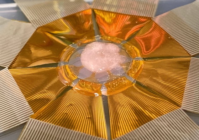
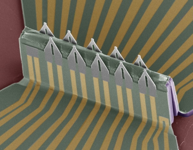
First author Yue Gu and colleagues present a more advanced approach in their study, involving a 3D array of highly sensitive field-effect transistors (FETs). These devices are shaped like sharp, pointed tips, and coated with a phospholipid bilayer: the membrane that forms a continuous boundary around all living cells. This allowed the sensors to non-invasively pierce through cell membranes, and detect electrical signals from directly inside.
To fabricate the device, the researchers first cut out the FETs in 2D shapes, then pinned them in specific positions to a pre-stretched elastomer sheet. When the sheet was then loosened, this material then buckled and bent into its final 3D shape.
Unlike previous patch clamping approaches, the team’s FET array could be used to monitor signals from multiple cells at the same time, and even at two different sites within the same cell. This enabled them to measure both the speeds and directions of signals within individual cells, as well as between specific pairs of cells within complex 3D networks.
To test their approach, the researchers studied the propagation of signals within heart muscle cell cultures and engineered 3D cardiac tissues, placed on top of the FET array. For the first time, this allowed them to measure intracellular signals in the 3D tissue. The experiment revealed some particularly intriguing behaviours: showing that electrical signals travel five times faster inside cells than between them.
The result could have broad implications for biologists’ understanding of cellular physiology. By identifying irregularities in signal propagations, the team hopes that clinicians could gain a detailed knowledge of heart disorders such as arrhythmia, heart attack and cardiac fibrosis. Although the practical medical use of the device is still some way off, the approach could eventually lead to FET arrays that can be implanted on real biological tissues, with artificial intelligence processing algorithms employed to offer valuable patient diagnoses.
The researchers describe their work in Nature Nanotechnology.
No hay comentarios:
Publicar un comentario