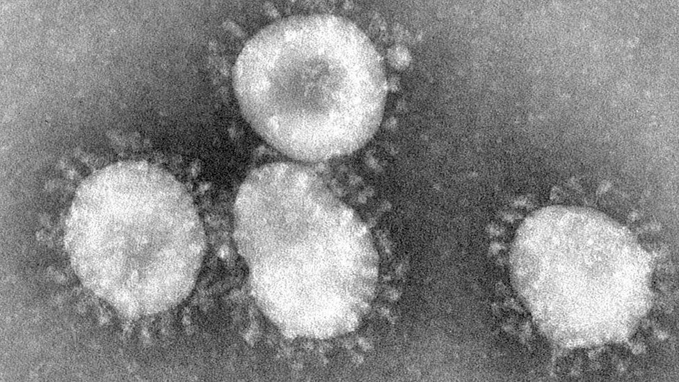
The woman who discovered the first coronavirus
The woman who discovered the first human coronavirus was the daughter of a Scottish bus driver, who left school at 16.
June Almeida went on to become a pioneer of
virus imaging, whose work has come roaring back into focus during the
present pandemic.
Covid-19 is a new illness but it is caused by
a coronavirus of the type first identified by Dr Almeida in 1964 at her
laboratory in St Thomas's Hospital in London.
The virologist was born June Hart in 1930 and grew up in a tenement near Alexandra Park in the north east of Glasgow.
She left school with little formal education
but got a job as a laboratory technician in histopathology at Glasgow
Royal Infirmary.
Later she moved to London to further her career and in 1954 married Enriques Almeida, a Venezuelan artist.
Common cold research
The couple and their young daughter moved to
Toronto in Canada and, according to medical writer George Winter, it was
at the Ontario Cancer Institute that Dr Almeida developed her
outstanding skills with an electron microscope.
She pioneered a method which better visualised viruses by using antibodies to aggregate them.
Mr Winter told
Drivetime on BBC Radio Scotland
her talents were recognised in the UK and she was lured back in 1964 to
work at St Thomas's Hospital Medical School in London, the same
hospital that treated Prime Minister Boris Johnson when he was suffering
from the Covid-19 virus.
On her return, she began to collaborate with
Dr David Tyrrell, who was running research at the common cold unit in
Salisbury in Wiltshire.
Mr Winter says Dr Tyrrell had been studying
nasal washings from volunteers and his team had found that they were
able to grow quite a few common cold-associated viruses but not all of
them.
One sample in particular, which became known
as B814, was from the nasal washings of a pupil at a boarding school in
Surrey in 1960.
They found that they were able to transmit
common cold symptoms to volunteers but they were unable to grow it in
routine cell culture.
However, volunteer studies demonstrated its
growth in organ cultures and Dr Tyrrell wondered if it could be seen by
an electron microscope.
They sent samples to June Almeida who saw the
virus particles in the specimens, which she described as like influenza
viruses but not exactly the same.
She identified what became known as the first human coronavirus.
Image copyright
Getty Images


Mr Winter says that Dr Almeida had actually
seen particles like this before while investigating mouse hepatitis and
infectious bronchitis of chickens.
However, he says her paper to a peer-reviewed
journal was rejected "because the referees said the images she produced
were just bad pictures of influenza virus particles".
The new discovery from strain B814 was
written up in the British Medical Journal in 1965 and the first
photographs of what she had seen were published in the Journal of
General Virology two years later.
According to Mr Winter, it was Dr Tyrrell and
Dr Almeida, along with Prof Tony Waterson, the man in charge at St
Thomas's, who named it coronavirus because of the crown or halo
surrounding it on the viral image.
Dr Almeida later worked at the Postgraduate Medical School in London, where she was awarded a doctorate.
She finished her career at the Wellcome Institute, where she was named on several patents in the field of imaging viruses.
After leaving Wellcome, Dr Almeida become a
yoga teacher but went back into virology in an advisory role in the late
1980s when she helped take novel pictures of the HIV virus.
June Almeida died in 2007, at the age of 77.
Now 13 years after her death she is finally
getting recognition she deserves as a pioneer whose work speeded up
understanding of the virus that is currently spreading throughout the
world.
-
https://www.bbc.com/news/amp/uk-scotland-52278716?fbclid=IwAR3fgkK4mCabop-5lC60nWeBJEbb9SchtF-6AMs-5LcgMFSfP8Il-L-hOiM
-
The Morphology of Three Previously Uncharacterized Human Respiratory Viruses that Grow in Organ Culture
Summary
A simple method is described for examining organ cultures by electron
microscopy for the presence of virus particles. The method was used to
detect the presence of three hitherto uncharacterized viruses. Two of
these have particles resembling those of infectious bronchitis of
chickens and the third morphologically resembles the parainfluenza group
of viruses.
- Received:
- Accepted:
- Published Online:
© Microbiology Society
Most read this month
-
https://www.microbiologyresearch.org/content/journal/jgv/10.1099/0022-1317-1-2-175?fbclid=IwAR37Psg14WaYSMOMbI5cpWlcPgE_LgYxOy2iOyNVulRn7sWzpRysAOzHBog#tab2
No hay comentarios:
Publicar un comentario